
A host of biologic and physiologic factors can shape long-term wound healing before a wound closes.1 This is especially important when an injury extends beyond the epidermis and into, or through, the dermis.
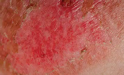
The epidermis has substantial regenerative capacity. Keratinocyte stem and progenitor cells located along the wound margins and within surviving skin appendages can restore the epithelial barrier. In contrast, extensive loss of the dermis, including its extracellular matrix, microvasculature, hair follicles, sebaceous glands, and other appendages, generally heals through repair rather than complete regeneration. The resulting scar restores continuity but does not fully reproduce the structure, elasticity, strength, or biological functions of uninjured skin.1,2
Although limited regenerative phenomena such as wound-induced hair-follicle neogenesis have been described, complete restoration of adult human dermis following a large full-thickness injury remains uncommon. Therefore, the clinically relevant goal is often to guide repair toward stable coverage, acceptable function, and the best possible scar outcome. A collagen-based wound matrix may support this process by providing a temporary three-dimensional scaffold within a properly prepared wound bed. The matrix does not heal the wound by itself. Rather, it creates a structure that host cells can enter, remodel, and gradually replace with newly formed tissue.
Collagen throughout the wound-healing cascade
Wound healing is commonly described as four overlapping phases: 1
Hemostasis → Inflammation → Proliferation → Remodeling
During hemostasis, bleeding is controlled and a provisional fibrin matrix forms. Inflammation follows as immune cells remove damaged tissue and coordinate the transition toward repair. During proliferation, fibroblasts deposit extracellular matrix, endothelial cells contribute to new blood-vessel formation, granulation tissue develops, and keratinocytes migrate across the wound surface. Finally, during remodeling, collagen is reorganized and cross-linked over months or longer, gradually increasing the strength of the repaired tissue.1,3
Collagen participates throughout this cascade. As a major component of the native extracellular matrix, it provides structural support and biological cues involved in cellular attachment, migration, signaling, and tissue organization.3,4 Fibroblast behavior and extracellular-matrix remodeling are particularly important because persistent activation of a fibrotic fibroblast phenotype can promote excessive collagen deposition and scar formation.5
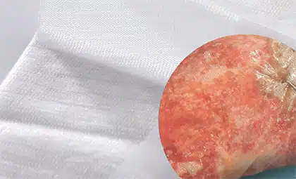
When the native dermal framework has been lost, a porous collagen matrix can provide a temporary structure into which host cells and blood vessels may enter as the implanted material is remodeled. 6 The objective is not to recreate normal dermis immediately, but to support organized vascularized tissue formation as part of a broader reconstructive strategy.
Not all wound matrices are the same
Wound matrices differ in source, composition, architecture, thickness, porosity, crosslinking, degradation rate, and handling characteristics. These differences can influence how a matrix interacts with the wound and how it fits within a reconstructive plan. 7
Biologic matrices may be derived from bovine, porcine, or human tissue. Synthetic matrices use manufactured polymers, while hybrid or biosynthetic products combine biological and synthetic components. These categories describe composition and not a universal hierarchy of clinical performance.
Architecture also matters.8 A single-layer matrix places the scaffold directly against the wound and is covered with an appropriate secondary dressing. A bilayer matrix generally combines a wound-contact scaffold with a temporary outer layer, often designed to help control moisture loss and protect the wound while the underlying matrix integrates. That outer layer may require removal before definitive closure.
Neither configuration is inherently appropriate for every wound. Matrix selection should consider:
- Wound depth and location
- Exposed structures
- Perfusion
- Exudate
- Contamination risk
- Available donor skin
- Timing of definitive closure
- Whether treatment is planned as a single- or multiple-stage reconstruction
A recent systematic review identified wound depth as a primary consideration in dermal-substitute selection while emphasizing that evidence-based indications continue to evolve.9
Successful application begins with the wound bed
Timing should be driven by wound biology rather than the calendar alone. A matrix cannot compensate for an inadequately prepared wound. Before application, clinicians should remove debris and nonviable tissue, achieve hemostasis, assess perfusion, and address infection according to the product labeling and the patient’s clinical needs. Off-loading, compression, nutritional support, glycemic management, and treatment of underlying vascular or systemic disease may be equally important, depending on wound etiology.
After placement, close contact between the matrix and wound bed must be maintained. Appropriate fixation, management of moisture and exudate, protection from shear, clinical surveillance, and selection of the appropriate time for definitive closure remain essential parts of the treatment pathway.
Do short-term effects predict long-term outcomes?
Matrix integration, vascularized tissue formation, readiness for grafting, graft take, complete closure, durable closure, scar quality, and function are related; however, they are distinct clinical outcomes.
A 2024 systematic review of dermal substitutes in burns and burn-scar reconstruction identified potential improvements in selected scar outcomes, but the studies varied considerably in the products, wounds, techniques, and outcomes evaluated. Few differences were consistently statistically significant.10
Similarly, a randomized within-patient study comparing a collagen-elastin dermal substitute plus split-thickness skin grafting against grafting alone found lower graft take and delayed healing in the matrix-treated areas. Although selected measurements of skin extension and elasticity were better at 12 months, other scar outcomes at 12 months and final scar quality at six years did not differ between treatments.11
Preclinical research evaluating a bovine dermal collagen matrix in porcine full-thickness wounds has demonstrated cellular infiltration, angiogenesis, persistent collagen scaffolding, and support of autografting at seven days.12 These findings help explain how matrix architecture may support wound-bed preparation but should not be interpreted as proof of equivalent long-term effects in patients.
The distinction is important: biological plausibility, matrix integration, or earlier graft readiness does not automatically establish superior long-term healing. A collagen-based matrix may help prepare a vascularized wound bed for closure; whether that translates into more durable closure, improved function, or better scar quality depends on the product, wound, patient, and clinical setting.
Collagen wound matrices: Key takeaways
Collagen-based wound matrices provide a temporary scaffold intended to support the body’s wound-repair process. Their clinical value depends on thoughtful matrix selection, thorough wound-bed preparation, appropriate application, and coordination of the complete reconstructive plan.
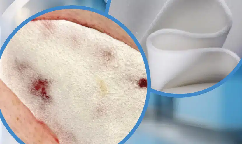
Cohealyx® collagen dermal matrix is a single-layer, porous, resorbable matrix engineered from purified bovine dermal collagen and indicated for the management of a range of wounds, including burns, as well as traumatic, surgical, and chronic wounds. Cohealyx should not be used on infected or contaminated wounds or in patients with a known allergy to bovine-derived collagen materials.13
Learn more about Cohealyx and consult the complete Instructions for Use for indications, contraindications, precautions, and application guidance.
References
- Peña OA, Martin P. Cellular and molecular mechanisms of skin wound healing. Nat Rev Mol Cell Biol. 2024;25(8):599–616. doi:10.1038/s41580-024-00715-1.
- Frech S, Lichtenberger BM. Modulating embryonic signaling pathways paves the way for regeneration in wound healing. Front Physiol. 2024;15:1367425. doi:10.3389/fphys.2024.1367425.
- Diller RB, Tabor AJ. The role of the extracellular matrix in wound healing: A review. Biomimetics. 2022;7(3):87. doi:10.3390/biomimetics7030087.
- Gardeazabal J, et al. Elastin and collagen fibres in cutaneous wound healing. Exp Dermatol. 2024;33(3). doi:10.1111/exd.15052.
- Jay JW, Palackic A, Prasai A, et al. Photoactivated rose bengal mitigates a fibrotic phenotype and improves cutaneous wound healing in full-thickness injuries. Wound Repair Regen. 2024;32(5):758–769. doi:10.1111/wrr.13202.
- Gupta S, Moiemen N, Fischer JP, Attinger C, Jeschke MG, Taupin P, Orgill DP. Dermal Regeneration Template in the Management and Reconstruction of Burn Injuries and Complex Wounds: A Review. Plast Reconstr Surg Glob Open. 2024 Mar 20;12(3):e5674. doi: 10.1097/GOX.0000000000005674. PMID: 38510326; PMCID: PMC10954069.
- Dilek ÖF, Sevim KZ, Dilek ON. Acellular dermal matrices in reconstructive surgery; history, current implications and future perspectives for surgeons. World J Clin Cases. 2024 Dec 16;12(35):6791-6807. doi: 10.12998/wjcc.v12.i35.6791. PMID: 39687641; PMCID: PMC11525903.
- Sharafi AM, Pakkhesal S, Monajjemzadeh F, Alipour N, Hamidi S. Preparation of Multilayer Platforms for Advanced Wound Care Management. Polymers (Basel). 2025 Sep 2;17(17):2393. doi: 10.3390/polym17172393. PMID: 40942311; PMCID: PMC12431120.
- van den Bosch AS, Verwilligen RAF, Pijpe A, et al. Indications for the use of dermal substitutes in patients with acute burns and in reconstructive surgery after burns: A systematic review. Wound Repair Regen. 2025;33(1). doi:10.1111/wrr.13248.
- van den Bosch AS, Verwilligen RAF, Pijpe A, et al. Outcomes of dermal substitutes in burns and burn scar reconstruction: A systematic review and meta-analysis. Wound Repair Regen. 2024;32(6):960–978. doi:10.1111/wrr.13226.
- Gardien KLM, Pijpe A, Brouwer KM, et al. Short- and long-term outcomes of an acellular dermal substitute versus standard of care in burns and reconstructions: A phase I/II intrapatient randomized controlled trial. Adv Skin Wound Care. 2023;36(10):540–548. doi:10.1097/ASW.0000000000000040.
- Bush KA, Nsiah BA, Jay JW, et al. Bovine dermal collagen matrix promotes vascularized tissue generation supporting early definitive closure in full-thickness wounds: A pre-clinical study. Cureus. 2025;17(3). doi:10.7759/cureus.81517.
- Cohealyx® Collagen Dermal Matrix. Instructions for Use. Rev. 0. February 2025.